Tungsten Filament Scanning Electron Microscope | SEM3200
Home > Products > Tungsten Filament Scanning Electron Microscope | SEM3200
CIQTEK SEM3200 is a high-performance tungsten filament scanning electron microscope. It has excellent image quality, low vacuum mode compatibility, high resolution images in different fields of view. The depth of field is large and the image is rich in stereo.
Accesories
- Autosampler AS-500 with optional rinse station Argon humidifier.
- Introduction system kits for improved performance (small volume, organics, Hydrofluoric acid, high total dissolved solids).
- Concomitant Metals Analyzer for simultaneous measurement of hydride forming elements and other elements.
- Oxygen kit for alkali elements in organics.
Extended Scalability and Functionalities
SEM SE\BSE\EDS\EDX\EBSD, etc
Optical Navigation
Quickly locate target samples and areas of interest
*Large-Scale Image Stitching
Fully automatic map picking and stitching to display a large field of view
Image Blending (SE+BSE)
Observe the composition and surface information of a sample in one image
*Dual Anode Structure
Improved resolution and imaging quality at low voltages
*Low Vacuum Mode
Provide sample surface details and morphology in low vacuum, software switch vacuum state with one click
Features
Low Voltage
- Carbon material samples with small penetration depth at low voltage. The real shape of the sample surface can be obtained with richer details.
- The electron beam irradiation damage of the hair sample is reduced at low voltage while the charge effect is eliminated.
Parameters
| Models | SEM3200A | SEM3200 | ||
|---|---|---|---|---|
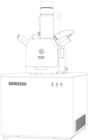
| Electro-Optical Systems | Electron Gun | Pre-aligned medium-sized fork-type tungsten filament | ||
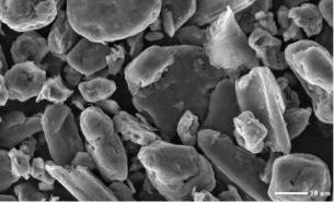
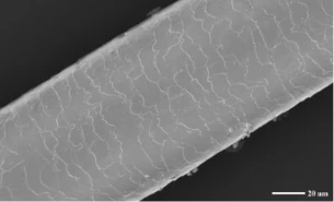
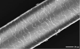
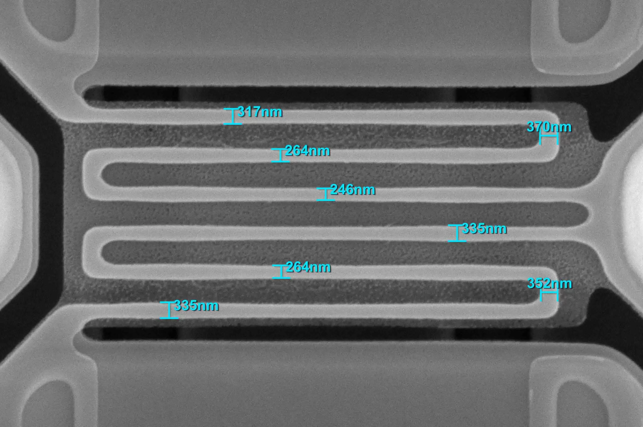
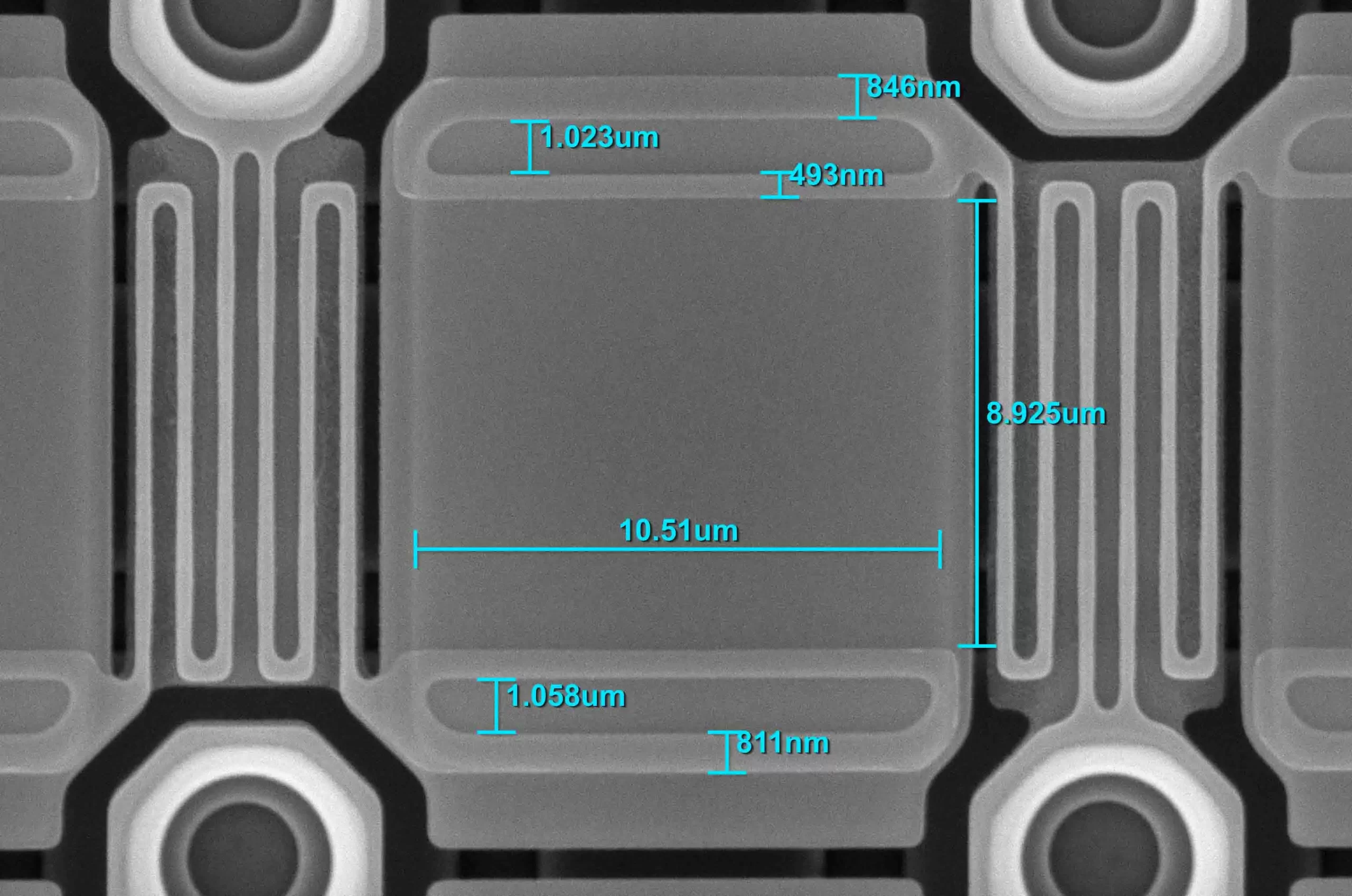
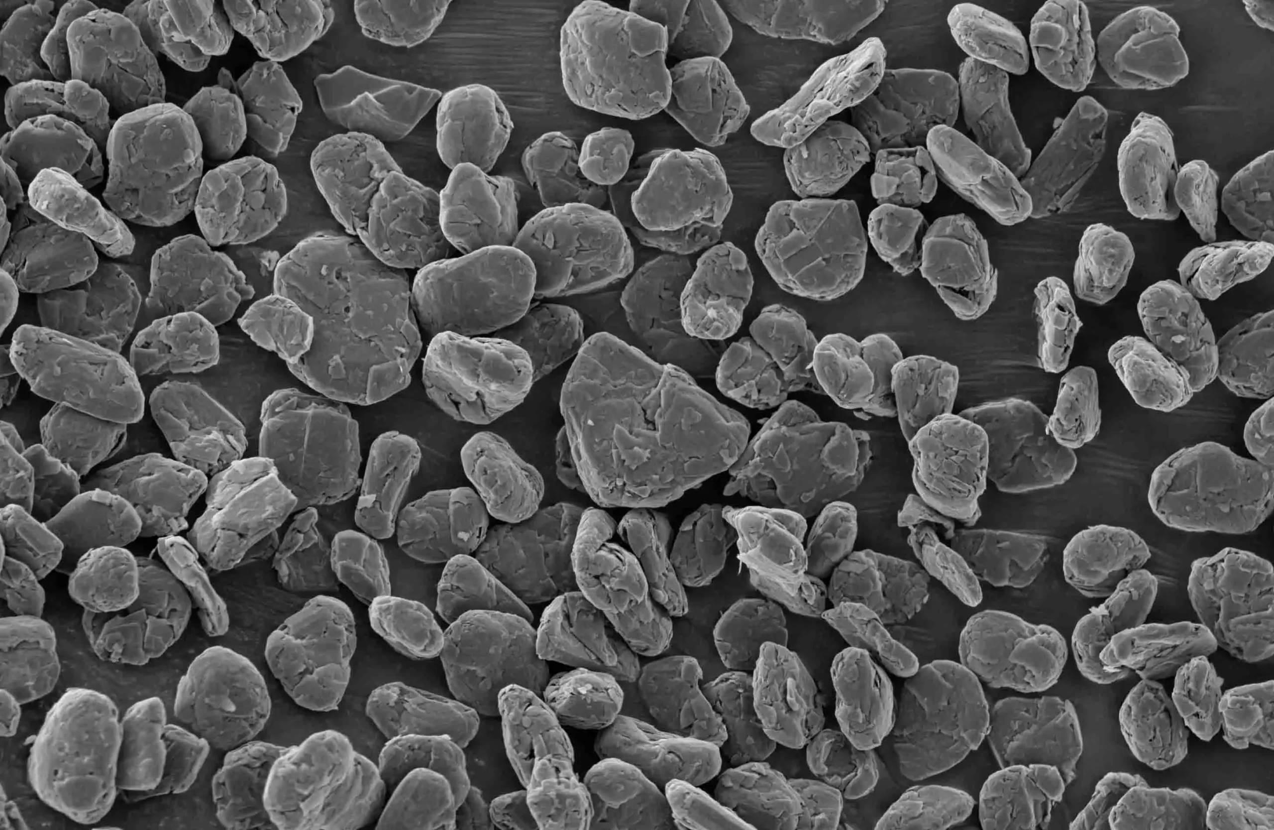
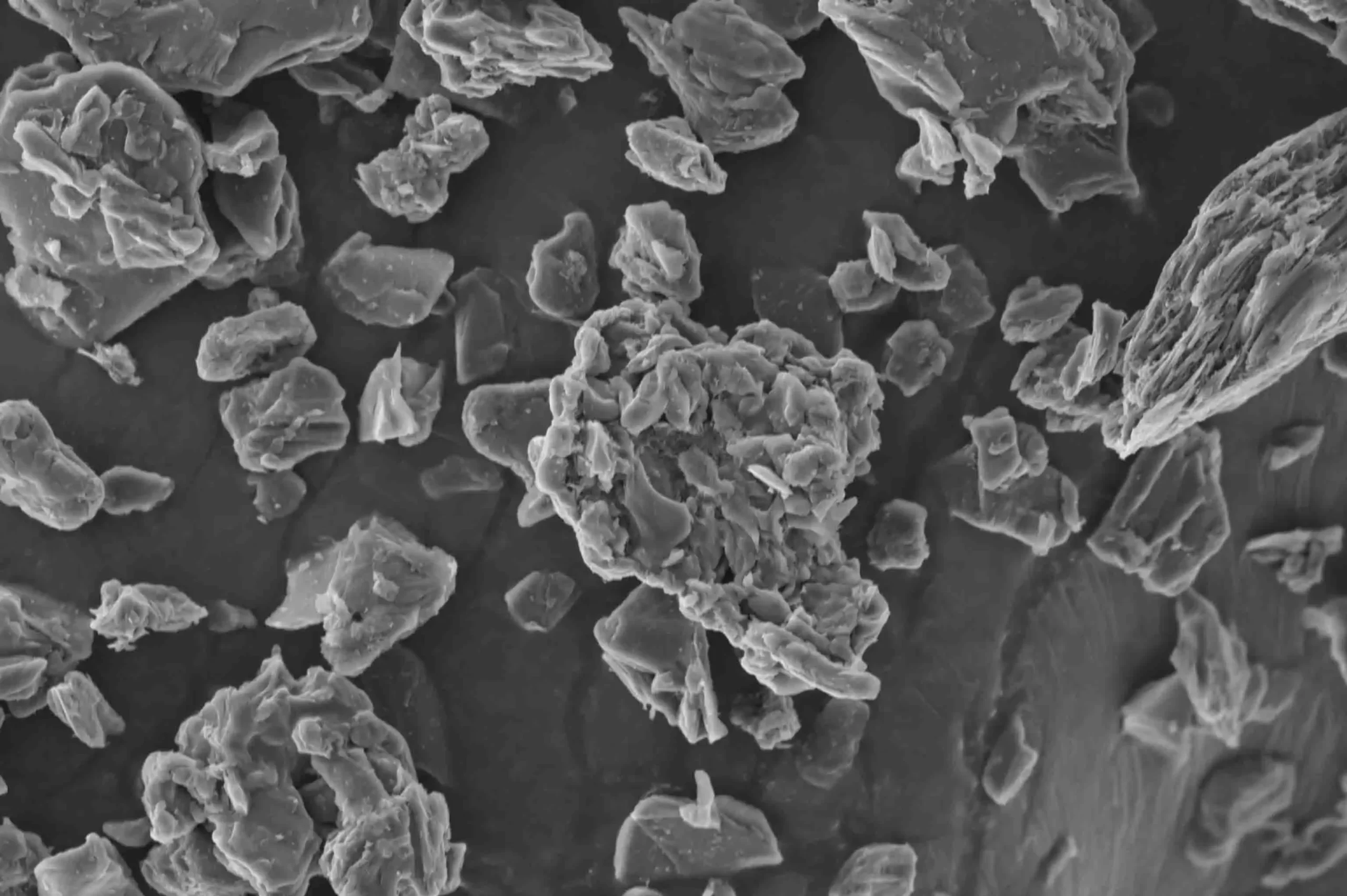
| Resolution | High Vaccum | 3 nm @ 30 kV (SE) | ||
| 4 nm @ 30 kV (BSE) | ||||
| 8 nm @ 3 kV (SE) | ||||
| *Low Vaccum | 3 nm @ 30 kV (SE) | |||
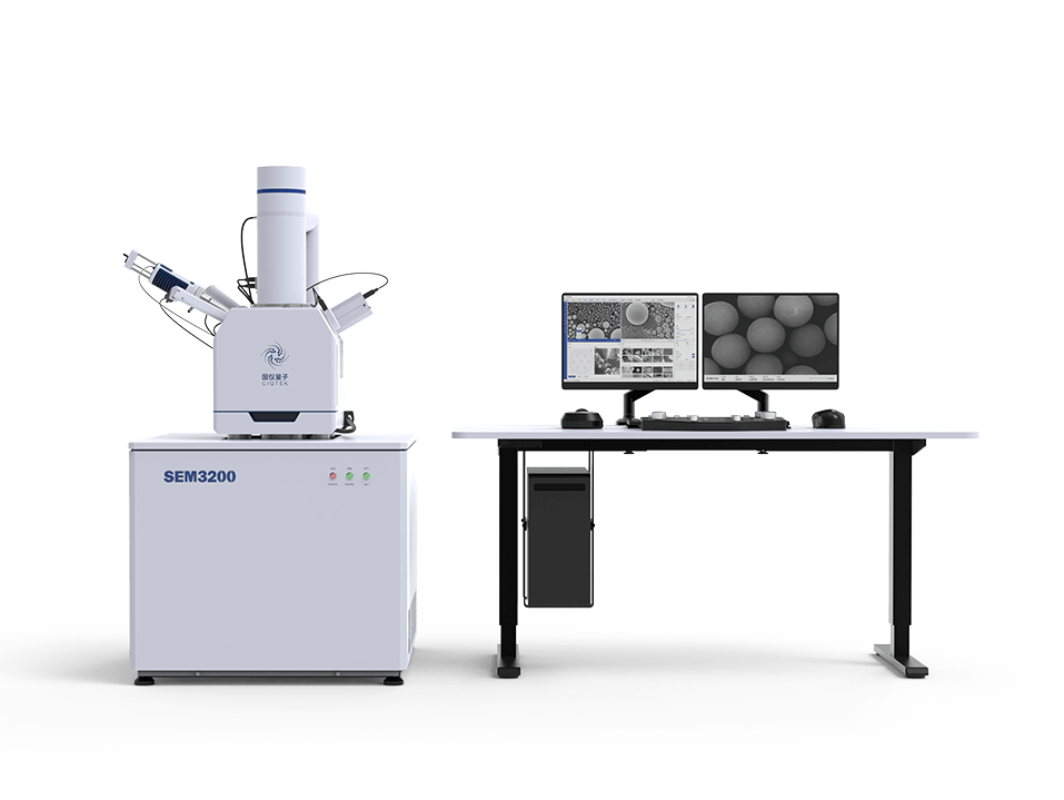
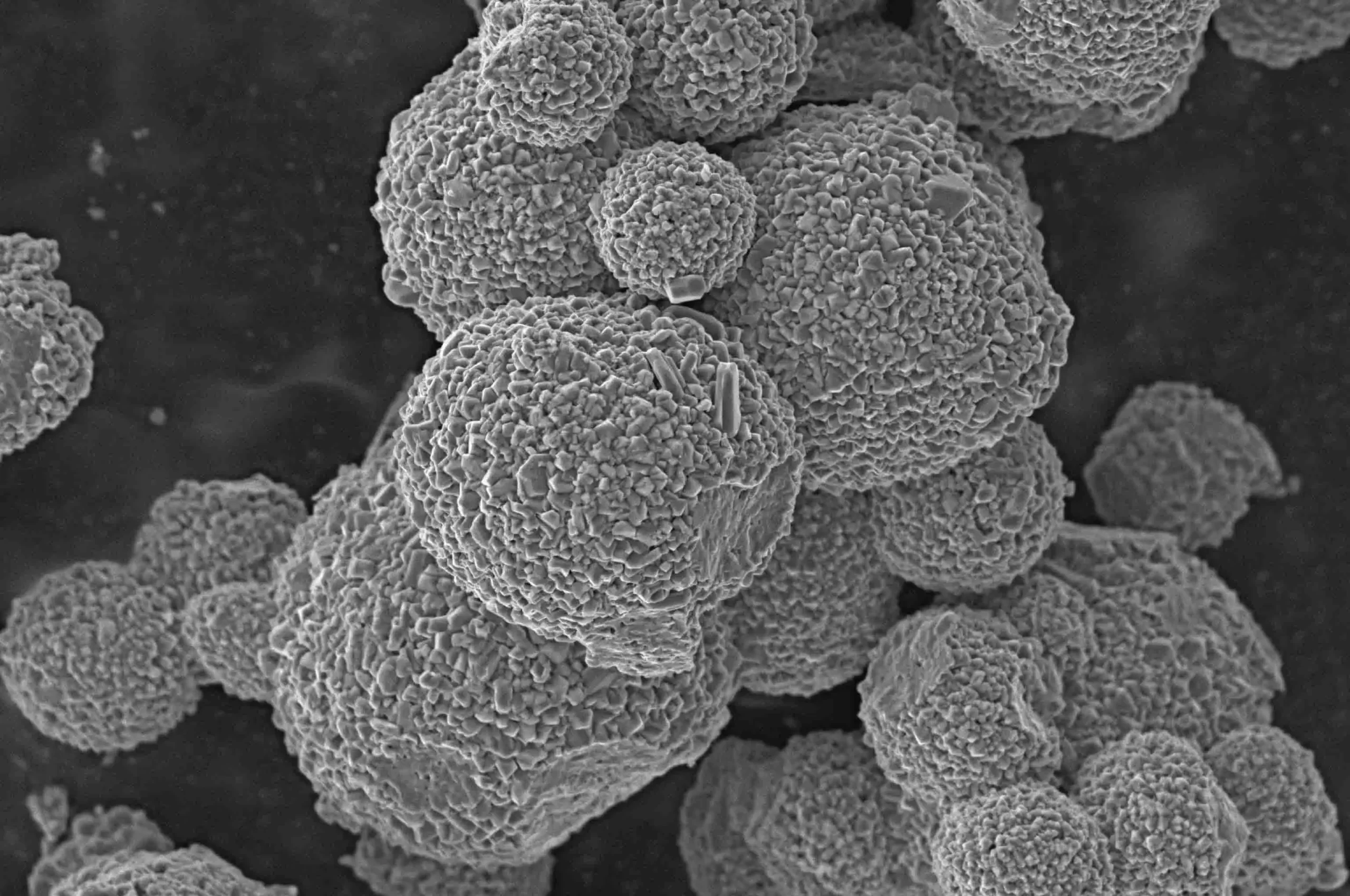
| Magnification | 1-300,000x (Film Magnification) | |||
| 1-1000,000x (Screen Magnification) | ||||
| Acceleration Voltage | 0.2 kV ~ 30 kV | |||
| Probe Current | ≥1.2μA, Real-time display | |||
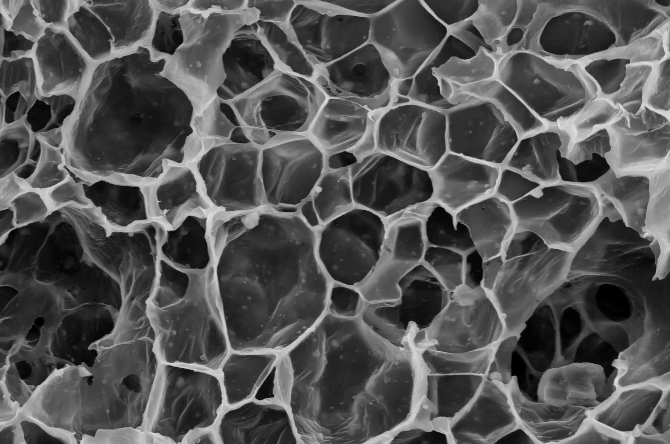
| Imaging Systems | Detector | Secondary Electron Detector (ETD) | ||
| *Backscattered electron detector (BSED), *low vacuum secondary electron detector, *energy spectrometer EDS, etc. | ||||
| Image Format | TIFF, JPG, BMP, PNG | |||
| Vacuum System | Vacuum Model | High Vacuum | Better than 5×10⁻⁴ Pa | |
| Low Vacuum | 5 ~ 1000 Pa | |||
| Control Mode | Fully automatic control | |||
| Sample Chamber | Camera | Optical Navigation | ||
| Monitoring in the Sample Chamber | ||||
| Sample Table | Three Axis Automatic | Five Axis Automatic | ||
| Distance | X: 120 mm | X: 120 mm | ||
| Y: 115 mm | Y: 115 mm | |||
| Z: 50 mm | Z: 50 mm | |||
| / | R: 360° | |||
| / | T: -10° ~ +90° | |||
| Software | Operating System | Windows | ||
| Navigations | Optical Navigation, Gesture Quick Navigation | |||
| Automatic Functions | Auto Brightness Contrast, Auto Focus, Automatic Dissipation | |||
| Special Functions | Intelligent Assisted Dispersion, *Large-Scale Image Stitching (Optional accessories) | |||
| Installation Requirements | Space | L≥ 3000 mm, W ≥ 4000 mm, H ≥ 2300 mm | ||
| Temperature | 20°C (68°F) ~ 25°C (77°F) | |||
| Humidity | ≤ 50 % | |||
| Power Supply | AC 220 V(±10 %), 50 Hz, 2 kVA | |||